Chondroblastic osteosarcoma of the maxilla: diagnostic challenges on incisional biopsy and report of a case
DOI:
https://doi.org/10.5935/2525-5711.20180012eKeywords:
Osteosarcoma, Chondrosarcoma, DiagnosisAbstract
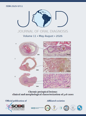
Osteosarcoma is a rare mesenchymal neoplasm characterized by osteoid production. Its neoplastic cells can synthesize variable kinds of matrix which determines the sub types as the chondroblastic variant and its cartilaginous matrix. We report a case of a 30-year- old man who presented a painful swelling in the right posterior maxilla, associated to teeth mobility. Radiographic exam showed bone rarefaction with poorly defined margins and root resorption in the corresponding area. An incisional biopsy was performed and histopathologic examination revealed proliferation of mesenchymal spindle-shaped cells in a stroma with irregular, immature and basophilic cartilaginous tissue without osteoid tissue. The cell were diffusely positive for S100 protein. A provisional diagnosis of chondrosarcoma was rendered until the excisional biopsy revealed the presence of osteoid tissue possibiliting a final diagnosis of chondroblastic osteosarcoma. Differentiating between chondrosarcoma and chondroblastic osteosarcoma in incisional biopsies may be a hard task when material is scarce, often incurring in the need for further biopsy. Correct diagnosis is essential for the correct treatment.
References
Raymond AK, Ayala AG, Knuutila S. Conventional Osteosarcoma. In: Fletcher CDM, Unni KK, Mertens F, eds. World Health Organization Classification of Tumours. Pathology and Genetics of Tumours of Soft Tissue and Bone. Lyon: IARC Press; 2002. p. 264-70.
Garrington GE, Scofield HH, Cornyn J, Hooker SP. Osteosarcoma of the jaws. Analysis of 56 cases. Cancer. 1967;20:377-91.
Bertoni F, Dallera P, Bacchini P, Marchetti C, Campobassi A. The Istituto Rizzoli-Beretta experience with osteosarcoma of the jaw. Cancer. 1991;68:1555-63.
Clark JL, Unni KK, Dahlin DC, Devine KD. Osteosarcoma of the jaw. Cancer. 1983;51:2311-6.
Unni K. Dahlin's Bone Tumors. General Aspects of Data on 11,087 Cases. 5th ed. Philadelphia: Lippincott-Raaven; 1996.
Paparella ML, Olvi LG, Brandizzi D, Keszler A, Santini-Araujo E, Cabrini RL. Osteosarcoma of the jaw: an analysis of a series of 74 cases. Histopathology. 2013;63:551-7.
McHugh JB, Thomas DG, Herman JM, Ray ME, Baker LH, Adsay NV, et al. Primary versus radiation-associated craniofacial osteosarcoma: Biologic and clinicopathologic comparisons. Cancer. 2006;107:554-62.
Patel SG, Meyers P, Huvos AG, Wolden S, Singh B, Shaha AR, et al. Improved outcomes in patients with osteogenic sarcoma of the head and neck. Cancer. 2002;95:1495-503.
Deshpande V, Nielsen GP, Rosenberg AE. Gnathic well differentiated osteosarcomas: A clinicopathologic study of 9 cases [abstract]. Modern Pathol. 2007:13A.
Desai D, Pandith S, Jeergal PA, Arathi K, Saini R. Fibroblastic variant of osteosarcoma: a challenge in diagnosis & management. Open Dent J. 2010,4:211-7.
Nakayama E, Sugiura K, Kobayashi I, Oobu K, Ishibashi H, Kanda S. The association between the computed tomography findings, histologic features, and outcome of osteosarcoma of the jaw. J Oral Maxillofac Surg. 2005;63:311-8.
Padilla RJ, Murrah VA. The spectrum of gnathic osteosarcoma: caveats for the clinician and the pathologist. Head Neck Pathol. 2011;5:92-9.
Inewards CY. Update on cartilage forming tumors of the head and neck. Head Neck Pathol. 2007;1:67-74.
Gomez-Brouchet A, Mourcin F, Gourraud PA, Bouvier C, De Pinieux G, Le Guelec S, et al. Galectin-1 is a powerful marker to distinguish chondroblastic osteosarcoma and conventional chondrosarcoma. Hum Pathol. 2010;41:1220-30.
Downloads
Published
How to Cite
Issue
Section
License
Copyright (c) 1969 Lara Cristina Oliver Gimenez, Bruno Tavares Sedassari, Cibele Pidorodeski Nagano, André Caroli Rocha, Suzana Cantanhede Orsini Machado de Sousa, Décio dos Santos Pinto-Júnior

This work is licensed under a Creative Commons Attribution 4.0 International License.